A 56-year-old man presents for evaluation of dyspnea on exertion. He has a 50
pack-year history of cigarette smoking and was hospitalized 5 years earlier
because of severe pneumonia. He has a daily cough without sputum production.
The chest is barrel-shaped, with prolonged expiration and diminished breath
sounds. Results of pulmonary function testing include the following: FEV1,
2.74 L (55% predicted); FVC, 5.48 L (89% predicted); FEV1/FVC, 50%;
TLC, 9.05 L (110% predicted); DLco, 19.73 ml/min/mm Hg (43%
predicted). After administration of albuterol, the FEV1 is 2.77 L
(56% predicted).
What is the most likely explanation for this patient's dyspnea?
Asthma
Bronchiectasis
Chronic bronchitis
Emphysema
Churg-Strauss vasculitis
Answer 1 is
not correct. Answer 4 is correct.
Key Concept: Recognize the pulmonary function test pattern of
emphysema
This patient has a reduced FEV1/FVC ratio and increased TLC,
consistent with obstructive lung disease. The most common causes of
obstructive lung disease are asthma, chronic bronchitis, and emphysema;
bronchiectasis is a less common cause. In emphysema, destruction of the
alveolar and capillary beds results in a reduced DLco. In pure
form, chronic bronchitis is characterized by chronic sputum production,
airflow obstruction, and minimal reduction in DLco. The absence of
bronchodilator response and reduced DLco are not consistent with a
diagnosis of asthma.
Question #14
A 30-year-old woman is brought to an emergency room for evaluation of a
reduced level of consciousness. Arterial blood gases measured while she was
breathing room air at sea level are pH, 7.16; Pco2, 70 mm Hg; Po2,
50 mm Hg.
What is the most likely explanation for this patient's hypoxia?
Diffusion abnormality
Hypoventilation
Intrapulmonary shunt
Ventilation-perfusion mismatch
Answer 1 is
not correct. Answer 2 is correct.
Key Concept: Understand how to use the alveolar gas equation to
distinguish among the causes of hypoxia
This patient's arterial blood gases demonstrate acute respiratory acidosis.
The alveolar-arterial oxygen gradient (A-aDo2), calculated using
the alveolar gas equation [the Pao2 = Pio2
- (Pao2 x 1.25)], is 12.5 mm Hg, which is a normal
result. Among the causes of hypoxia listed, only hypoventilation would not
affect the A-aDo2.
Question #15
You are asked to assess a critically ill patient with adult respiratory
distress syndrome and multiple organ failure. The blood pressure is 100/70,
and heart rate is 100. While the patient is undergoing mechanical ventilation
with an inspired oxygen concentration of 60%, the Pao2
is 62 mm Hg (oxygen saturation, 90%). A pulmonary arterial catheter is in
place; the pulmonary arterial occlusion pressure is 3 mm Hg, and the cardiac
output is 9 L/min (normal, 4-8 L/min). The hematocrit is 26%, and the
hemoglobin is 6 g/dl.
Which of the following interventions would be the most effective way to
increase tissue oxygen delivery in this patient?
Transfusion of packed red blood cells to increase hemoglobin to 9
g/dl
Addition of positive end-expiratory pressure to increase oxygen
saturation to 96%
Dobutamine infusion to increase cardiac output to 10 L/min
Crystalloid infusion to increase pulmonary arterial occlusion pressure
Answer 1 is
correct.
Key Concept: Understand the principles of systemic oxygen transport
The total amount of oxygen delivered to the systemic circulation is the
product of the cardiac output and the arterial content of oxygen per unit of
arterial blood (Cao2). The Cao2 is
determined by the concentration and characteristics of hemoglobin and the
arterial oxygen saturation [Cao22 = Hb (g/dl) x Sao2
(%) x 1.39 ml O2/g Hb]. In this patient, increasing the hemoglobin
concentration by 3 g/dl would increase arterial oxygen delivery more than
would a 6% rise in oxygen saturation or a 1 L/min increase in cardiac output
(with normal cardiac function, dobutamine would be contraindicated). Infusion
of crystalloid to raise the pulmonary arterial occlusion pressure to the
normal range would be a reasonable intervention but would not be expected to
increase the cardiac output significantly.
Question #16
A colleague asks you to interpret the following pulmonary function test
results: FEV1, 3.08 L (50% predicted); FEV1/FVC, 85%;
TLC, 4.30 L (52% predicted); RV, 1.64 L (75% predicted); DLco, 44.5
ml/min/mm Hg (95% predicted).
Which of the following would you suggest?
Further workup for pulmonary vascular disease
Trial of bronchodilators
Assess chest wall and neuromuscular status
High-resolution CT scan to evaluate interstitial lung disease
Ventilation-perfusion scan
Answer 1 is
not correct. Answer 3 is correct.
Key Concept: Recognize the restrictive pulmonary function test pattern
of chest wall and neuromuscular disorders
The pulmonary function test results reveal a symmetrical reduction in FEV1
and TLC, with preserved FEV1/FVC ratio, a restrictive pattern.
Restrictive defects can be caused by interstitial lung diseases, chest wall
disorders, and neuromuscular disorders. Of the choices given, chest wall and
neuromuscular disorders would produce a restrictive pattern with normal DLco.
Interstitial lung diseases of sufficient severity to reduce lung volumes by
50% would generally result in destruction of the lung parenchyma and a lower
DLco. A normal DLco is not consistent with a diagnosis
of pulmonary vascular disease. Bronchodilators would not be indicated in the
absence of airflow obstruction.
Question #17
A 45-year-old man presents with dyspnea. Three months earlier he required
prolonged endotracheal intubation during treatment for Guillain-Barre
syndrome, from which he has otherwise recovered. Fiberoptic bronchoscopy
demonstrates tracheal stenosis 5 cm above the carina.
Which of the following flow-volume loop patterns would be expected to result
from this complication?
Normal inspiratory flow rate with reduced expiratory flow rate
Reduced inspiratory and expiratory flow rates
Reduced inspiratory flow rate with normal expiratory flow rate
No effect on inspiratory and expiratory flow rates
Answer 1 is
not correct. Answer 2 is correct.
Key Concept: Understand flow-volume loop patterns
This patient presents with tracheal stenosis, a rare complication of prolonged
endotracheal intubation. The result would be a fixed obstruction of airflow
during inspiration and expiration. Variable airflow obstruction occurs when
the tracheal wall is weak. In such disorders, airflow rates are determined by
the location of the tracheal abnormality and the relationship between
intraluminal airway pressure and the pressure of the surrounding tissues. In
intrathoracic tracheomalacia, expiratory flow is limited because intrathoracic
pressure exceeds that within the trachea, the lumen narrows, and flow is
obstructed. When the abnormality occurs in the extrathoracic trachea (vocal
cord paralysis and extrathoracic tracheomalacia are examples), inspiratory
flow is reduced because the pressure within the extrathoracic upper airway
during inspiration is less than the surrounding tissue pressure because of the
Bernoulli effect.
Question #20
A 65-year-old woman is admitted for treatment of a community-acquired
pneumonia. On admission, her respiratory rate is 28.
Which factor may contribute to this patient's dyspnea?
Hypothermia
Decreased input from the carotid or aortic bodies
Rise in cerebrospinal fluid pH
Activation of pulmonary C fibers
Fall in arterial Pco2
Answer 1 is
not correct. Answer 4 is correct.
Key Concept: Know the physiologic mechanisms responsible for
ventilatory control
Activation of pulmonary C fibers will produce an increase in the ventilatory
drive. These are unmyelinated fibers whose afferent signal travels through the
pulmonary branch of the vagus nerves. They are probably responsible for the
tachypnea seen with such lung disorders as pulmonary edema, pneumonia, ARDS,
and interstitial disease. Hypothermia decreases ventilatory drive by reducing
metabolic rate, thereby decreasing production of CO2. This, in
turn, will lower arterial Pco2, which will raise cerebrospinal
fluid pH. These changes are sensed by chemoreceptors situated on the surface
of the medulla, which responds by lowering ventilatory drive. Impulses from
the carotid bodies are triggered by decrease in arterial Pco2;
these impulses travel via the ninth and tenth cranial nerves to the medullary
respiratory centers, where they stimulate increases in tidal volume and
breathing rate.
Question #21
A 70-year-old man is admitted to your service for exacerbation of his
congestive heart failure. His other past medical history is significant for
end-stage renal disease secondary to diabetes, now requiring hemodialysis. On
rounds the day after admission, he is somnolent with a breathing pattern that
steadily increases and then decreases in size in a smooth
crescendo-decrescendo pattern, followed by apneas.
This patient's respiratory pattern would NOT be seen in which of the following
conditions?
Severe congestive heart failure
Hypoxia
Hemodialysis
Sleep
Pontine hemorrhage
Answer 1 is
not correct. Answer 5 is correct.
Key Concept: Recognize and understand the pathophysiology of Cheyne-Stokes
breathing
Cheyne-Stokes breathing can be seen with lesions deep in the cerebral
hemispheres and basal ganglia (such as infarctions or hypertensive
encephalopathy), not pontine lesions, which produce apneustic breathing. Other
than these CNS structural causes, Cheyne-Stokes breathing is produced by
alteration in CO2 homeostasis, usually hypocapnia. Hypoxia (caused
by high altitude, as well as other conditions) stimulates the respiratory
drive, lowering Pco2, thereby lowering the respiratory drive.
Overshooting then results in hypoxia, setting up an oscillating pattern.
Congestive heart failure produces the same oscillation, thought to be caused
by the delay in circulatory time from the pulmonary capillaries to
chemoreceptor sites. A ventilatory disturbance may initiate an appropriate
change in ventilation from the controller, but because of the long circulation
time, the alterations in Paco2 and Pao2
caused by this change in ventilation will not be sensed by chemoreceptor sites
until an overcompensation by the breathing apparatus has occurred. Then, a
change in ventilation in the opposite direction occurs, again with overshoot,
and an oscillating breathing pattern is established. Hemodialysis can cause
the same problem by removing CO2 sufficiently to lower the Paco2,
thus depressing rhythmic activity of medullary respiratory neurons, producing
apneas and periodic breathing.
Question #22
A 60-year-old woman presents for evaluation of excessive daytime sleepiness.
She reports that she has always snored and that her husband has begun to
notice increased sleep movements and times when she stops breathing. Her
tiredness has increased to the point that she is falling asleep at her desk.
Her habits are remarkable for three to four alcoholic drinks per night. On
physical exam, you note obesity, hypertension, a "crowded" posterior
pharynx, bibasilar inspiratory rales, and dependent edema.
What is the next most appropriate diagnostic test for this patient?
Echocardiogram
Chest x-ray
Home nocturnal oxygen saturation monitoring
Full polysomnographic tests in a sleep laboratory
Thyroid stimulating hormone
Answer 1 is
not correct. Answer 4 is correct.
Key Concept: Recognize and order the appropriate diagnostic test for
obstructive sleep apnea syndrome (OSAS)
Several features of this case suggest OSAS. During sleep, there is a decrease
in tone in the upper airway that, combined with recumbency, results in
collapse of the upper airway with partial or total obstruction; partial
obstruction may become total when inspiratory effort produces suction that
collapses the airway. Increased tonic activity of the pharyngeal muscles may
try to compensate for this, but it is ineffectual in people with OSAS.
Although OSAS tends to affect more men than women, with an estimated 24% of
men and 9% of women affected, the prevalence among women goes up after
menopause. Alcohol exaggerates relaxation of the upper airway muscles,
worsening the obstruction. Obesity may contribute to the problem because of
deposition of fatty tissue along the walls of the hypopharynx, as may
structural abnormalities such as midface hypoplasia, micrognathia or
retrognathia, macroglossia, nasal obstruction, and enlargement of the tonsils
or adenoids (though in most adults these are not present). The best diagnostic
test is full polysomnography in a sleep lab. Home nocturnal oxygen saturation
monitoring with evidence of desaturation may suggest the diagnosis but is not
confirmatory.
Question #23
For the patient in Question 22, you proceed with full polysomnographic tests
in a sleep laboratory, which demonstrate repetitive episodes of desaturation,
frequent apneas, and sleep disruptions occurring 60 to 70 times per hour; the
diagnosis of OSAS is made. Potential consequences of this condition include
which of the following?
Increased risk of automobile accidents
Hypertension
Ischemic heart disease
Right-sided heart failure
All of the above
Answer 1 is
not correct. Answer 5 is correct.
Key Concept: Know the consequences of OSAS
OSAS produces myriad health problems. People with this diagnosis carry a
sevenfold increased risk of automobile accidents, largely resulting from
somnolence caused by sleep disruptions from frequent apneas (though the
frequency of sleep disruptions correlates only loosely with the degree of
somnolence). Physicians should inform patients of this increased risk and may
be required to report patients who go untreated. Hypertension is brought on by
increased catecholamine release, induced by frequent hypoxic events. Ischemic
heart disease is promoted by hypertension (as above), elevation in serum
lipids and catecholamines, and heightened platelet activation, all of which
can be seen in moderate to severe OSAS. Nocturnal hypoxemia and hypercapnia
cause pulmonary arterial vasoconstriction that increases pulmonary pressures
and can lead to right-sided heart failure.
Question #24
What is the most effective therapy for the patient in Question 22?
Weight loss
Nocturnal continuous positive airway pressure (CPAP) by nasal mask
Medroxyprogesterone
Uvulopalatopharyngoplasty
Acetazolamide
Answer 1 is
not correct. Answer 2 is correct.
Key Concept: Know the treatments for OSAS
Nasal CPAP is the most effective and frequently applied therapy for OSAS. It
provides a pneumatic splint that prevents inspiratory collapse of the upper
airway during all phases of sleep when the patient is in the supine position.
The therapeutic results are often dramatic, with prompt resolution of
fragmented sleep and daytime sleepiness. Many of the sequelae of OSAS are
reversible as well. However, it is effective only when used regularly, and
some patients have difficulty with compliance. Weight loss is an important
adjunct in the treatment of OSAS, but long-term weight reduction is usually
difficult to maintain. Medroxyprogesterone can be effective in nonobstructive
sleep apnea, but it is a second-line therapy for OSAS and should be reserved
for patients who do not respond to nocturnal CPAP. Acetazolamide can prevent
hypocapnia-induced apnea, but there are no data on its long-term efficacy.
Surgical therapies can be useful in carefully selected patients with
well-defined anatomic anomalies, but these are the minority, and again, CPAP
is the first-line therapy.
Respiratory Medicine
Question #13
A 70-year-old man with severe ankylosing spondylitis presents with complaint
of shortness of breath. He denies fever or cough but does note mild right
lateral chest pain that seems to worsen with inspiration. His wife reports
that several months ago, he had a small stroke, which resulted in moderate
residual right hand and right leg weakness; the patient requires some
assistance with daily activities. Physical exam is remarkable only for mild
tachypnea (22/min); breath sounds are normal. Chest radiograph shows mild
hyperinflation, which is unchanged from previous studies. The ECG is normal,
as are serum myoglobin and creatinine kinase levels. Arterial blood gas shows
a pH of 7.32, a Pco2 of䀠31, and a Po2 of 80 (room
air).
What is the most appropriate next step in managing this patient?
Ventilation-perfusion scanning to evaluate for pulmonary embolism
Sputum and blood cultures, followed by broad-spectrum antibiotics to
treat early pneumonia
Spirometry to evaluate for worsening restriction caused by ankylosing
spondylitis
Serial troponin I assay to evaluate for myocardial infarction
Cardiac echocardiography to detect possible left ventricular dysfunction
Answer 1 is
correct.
Key Concept: Recognize pulmonary embolism in a patient with chest wall
disease
This patient has an elevated alveolar-arterial oxygen gradient that is
unexplained by his underlying chest wall disorder and findings on chest x-ray.
His previous stroke and the resultant decrease in mobility place him at risk
for pulmonary embolism, a potentially fatal cause of hypoxemia that needs to
be evaluated.
The absence of signs and symptoms of infection together with a negative chest
x-ray make empiric antibiotic therapy inappropriate. Ankylosing spondylitis
may produce a reduction in vital capacity and total lung volume but will not
produce an elevation of the alveolar-arterial oxygen gradient. Myocardial
ischemia is a possibility; however, the pleuritic nature of this patient's
chest pain and the elevated alveolar-arterial pressure gradient make this
diagnosis less likely. Similarly, the clinical presentation is atypical for
congestive heart failure.
Question #14
A 57-year-old woman with a history of moderate chronic obstructive pulmonary
disease (COPD) and tobacco use presents with 3 days of worsening cough and
shortness of breath. Her sputum, which is normally white, is now thick and
yellow. Physical exam is significant for scattered rhonchi and wheezes.
Arterial oxygen saturation is 92%, which is unchanged from previous office
measurements. Chest x-ray shows mildly increased interstitial markings without
focal opacities and a significantly elevated left hemidiaphragm. She is
treated with a 5-day course of corticosteroids and p.r.n. albuterol, which
result in her returning to her usual state of health.
What is the most appropriate follow-up for this patient?
Spirometry with total lung volumes to establish baseline pulmonary
function
Arterial blood gas on room air to determine if she is chronically
retaining CO2
Follow-up chest x-ray in 1 month to ensure normalization
Chest CT to evaluate the cause of her diaphragm paralysis
This patient's presentation is most consistent with an uncomplicated
exacerbation of her COPD. The elevated hemidiaphragm, however, indicates the
possibility of unilateral diaphragmatic paralysis, a process that is probably
unrelated to her acute presentation. Neoplastic invasion of the phrenic nerve
is the most common cause of this and should be investigated, especially in
this middle-aged smoker.
Question #15
A 37-year-old woman with myasthenia gravis presents with cough, fever, and
shortness of breath of 2 days' duration. Arterial blood gas on room air shows
a pH of 7.42, a Pco2 of 34, and a Po2 of 80. Chest x-ray
shows right middle and right lower lobe consolidation. Gram's stain shows 3+
gram-negative rods and 4+ white blood cells. She is treated empirically with
ceftazidime, gentamicin, supplemental oxygen, and aggressive respiratory
therapy. During the next 24 hours, she becomes increasingly tachypneic and
short of breath. With the patient receiving supplemental oxygen (4 L/min),
repeat blood gas measurements show a pH of 7.31, a Pco2 of 49, and
a Po2 of 78.
What is the most appropriate therapy for this patient?
Add vancomycin to the antibiotic regimen
Discontinue gentamicin and start ciprofloxacin
Discontinue ceftazidime and start imipenem/cilastatin
Institute noninvasive positive pressure ventilation
Continue current therapy
Answer 1 is
not correct. Answer 2 is correct.
Key Concept: Recognize gentamicin as a weak neuromuscular blocking
agent
Gentamicin is a weak neuromuscular blocking agent that can cause significant
weakness in patients with underlying myasthenia. Other commonly used drugs
that may produce similar problems include clindamycin, propranolol, lincomycin,
streptomycin, and neomycin. Electrolyte abnormalities such as hypokalemia,
hypophosphatemia, and hypocalcemia may compound this effect. This woman's
arterial gases show progressive hypoventilation, consistent with respiratory
muscle weakness. Although this may be caused by respiratory muscle fatigue
secondary to myasthenia gravis, the gentamicin should be discontinued to
eliminate any possible contribution.
Question #16
A 27-year-old man with complete C4 quadriplegia after a motor vehicle accident
2 months ago reports significant shortness of breath. His symptoms have been
present for 3 to 4 weeks. He has not noted any congestion, increased
secretions, fever, or chills. He is able to sleep comfortably at night but
notes significant shortness of breath during the day. Physical exam is notable
for moderate obesity and complete paralysis below C4.
Which of the following is most likely to improve this patient's symptoms?
Assisted coughing every 4 hours
Noninvasive positive pressure ventilation via nasal mask during the day
Ipratropium bromide nebulization treatments every 6 hours
Diaphragmatic strengthening exercises
Abdominal support binder during the day
Answer 1 is
not correct. Answer 5 is correct.
Key Concept: Recognize platypnea associated with high cervical
quadriplegia
Quadriplegic patients lack abdominal muscle tone; as a result, when
quadriplegic patients assume an upright position, the weight of the abdominal
contents pulls the diaphragm down to a resting position that is lower than
normal. This results in smaller diaphragmatic excursions during inhalation and
smaller tidal volumes. These results can be easily assessed by upright and
supine spirometry. The diagnosis is supported by the finding of a decrease in
vital capacity when the patient is in an upright position. Properly fitted
abdominal binders provide abdominal support sufficient to prevent symptomatic
platypnea.
Question #17
A 43-year-old man is seen in the emergency room with cough and fever. A chest
x-ray is obtained and shows no evidence of pneumonia. A single small nodule is
noted in the right upper lobe. The nodule is approximately 8 mm in diameter
and contains small specks of calcium. The patient does not recall ever having
had a chest x-ray, and he does not have a primary care provider. The emergency
room physician refers this patient to a general internist for follow-up.
What is the best course of action at this time?
Repeat chest x-ray in 6 months
Repeat chest x-ray in 1 year
Thin-section CT with contrast enhancement
Refer patient for fine-needle biopsy
On the basis of the calcification pattern, the nodule may be considered
benign
Answer
3 is correct.
Key Concept: Understand the appropriate evaluation of single small
lung nodules
Thin-section CT with contrast enhancement is a new way to prove that a nodule
is benign. Patients younger than 35 years with pulmonary nodules may undergo
serial follow-up for 2 years. This patient is 43 years of age, and so the
nodule must be assumed to be malignant until it is proved benign. The nodule
may be assumed to be benign if a chest x-ray taken 2 or more years earlier
shows the lesion to be either the same size or larger than its current size.
This patient did not have any previous chest x-rays. It is inappropriate to
simply follow a nodule unless the patient is younger than 35 years. Although
fine-needle biopsy may provide a diagnosis by histopathology, by cytologic
exam, or by culture of a microorganism, it should not be performed until after
the less invasive chest CT scan in this case. Small specks of calcium and
dystrophic calcium are often seen in malignant tumors.
Question #18
A 56-year-old public health nurse who lives in Arizona is seen in clinic with
a cough of 1 month's duration. Her medical history is notable for systemic
lupus erythematosus (SLE); breast cancer, for which she underwent modified
radical mastectomy of the right breast 8 years ago without recurrence; and
moderate mitral valve insufficiency. A chest x-ray reveals multiple pulmonary
nodules of varying size scattered throughout both lungs.
What is the most likely cause of this patient's pulmonary nodules?
Tuberculosis
Metastatic breast cancer
Pulmonary nodules secondary to SLE
Endocarditis
Coccidioidomycosis
Answer
2 is correct.
Key Concept: Recognize the most likely cause of multiple pulmonary
nodules
Hematogenous metastases are the most likely cause of multiple pulmonary
nodules, especially if the patient is not febrile and the nodules vary in
size. This patient does have potential tuberculosis exposure. Primary
tuberculosis is less likely in this patient, given her age and the possibility
of ongoing tuberculosis exposure. Primary tuberculosis is often focal with
ipsilateral hilar adenopathy. Reactivation tuberculosis is often multifocal
with bilateral infiltrates in the upper lung zones. SLE may result in an
associated pneumonitis that may appear diffuse or as dense lower lobe
infiltrates. Pyemic abscesses may appear as multiple small nodules. These are
more likely to be associated with right-sided endocarditis involving the
tricuspid valve. Coccidioidomycosis can produce scattered nodules. The disease
is relatively inactive, and the only clinical finding may be an abnormal chest
x-ray. This patient could have incidental coccidioidomycosis, but metastatic
carcinoma is more likely.
Question #19
A previously healthy 38-year-old woman is seen in clinic with a fever and a
cough that is productive of purulent sputum. She notes that she developed
shaking chills 1 day ago and feels increasingly short of breath. A complete
blood count reveals a white blood count of 25,000, with a shift to the left.
Chest x-ray reveals a dense focal pulmonary infiltrate.
What is the most likely cause of this patient's pneumonia?
Mycoplasma pneumoniae
Legionella pneumophila
Staphylococcus aureus
Haemophilus influenzae
Streptococcus pneumoniae
Answer
5 is correct.
Key Concept: Recognize the most common cause of focal infiltrates
The most common cause of a focal infiltrate is bacterial pneumonia. Streptococcus
pneumoniae is the most common cause of bacterial pneumonia, accounting for
perhaps 85% of all cases in healthy young adults. Haemophilus influenzae,
Staphylococcus aureus, and Legionella pneumophila are associated
more frequently with bacterial pneumonia in patients with chronic medical
conditions and advanced age. Mycoplasma pneumoniae is associated with
focal infiltrates and would be another possible cause in this patient, but it
is a less likely cause than Streptococcus pneumoniae.
Question #20
A 53-year-old woman presents with a protracted course of malaise, weight loss,
and nonproductive cough. She notes that 2 months ago, before she experienced
her current symptoms, she had a flulike illness. Multiple dense peripheral
pulmonary infiltrates are noted on chest x-ray. After transbronchial lung
biopsy, a diagnosis of bronchiolitis obliterans organizing pneumonia (BOOP) is
established.
Which of the following is characteristic of BOOP?
BOOP responds better to glucocorticoid therapy than does idiopathic
pulmonary fibrosis
Most cases are associated with specific immunologic disorders
Pathologic findings are specific
Most patients require open lung biopsy to establish the diagnosis
The extent of disease is determined by chest x-ray
Answer 1 is
correct.
Key Concept: Recognize the key features of BOOP
Idiopathic BOOP is a chronic parenchymal lung disease that is clinically
distinct from idiopathic pulmonary fibrosis; it responds better to
glucocorticoid therapy and has a better prognosis than the latter condition.
Fully two thirds of patients respond dramatically to glucocorticoid therapy.
CT scanning shows the extent of disease better than chest radiography.
Features include patchy bilateral air-space consolidation, small nodular
opacities, bronchial wall thickening, and dilatation. Diagnosis can be made on
transbronchial lung biopsy. A minority of patients require open lung biopsy to
establish the diagnosis. Pathologic findings are nonspecific and may occur in
viral infections, toxic exposures, and hypersensitivity pneumonitis. Perhaps
one half of all cases are idiopathic.
Question #21
A 53-year-old man presents to clinic with fever, chills, and a productive
cough. He has a long history of atopic asthma. Allergic bronchopulmonary
asthma is suspected.
Which of the following is a pathognomonic finding in allergic bronchopulmonary
asthma?
Eosinophil count higher than 10,000/mm3
Branching fingerlike shadows of dilated central bronchi seen on chest
x-ray
Positive sputum cultures for Aspergillus
Positive skin tests to Aspergillus antigens
Hyphae seen on microscopic exam of sputum
Answer 1 is
not correct. Answer 2 is correct.
Key Concept: Recognize pathognomonic radiologic features of allergic
bronchopulmonary aspergillosis
Branching fingerlike shadows from mucoid impaction of dilated central bronchi
are virtually pathognomonic of allergic bronchopulmonary aspergillosis.
Eosinophil counts greater than 10,000/mm3 are often seen in Churg-Strauss
syndrome. Although the other choices are associated with allergic
bronchopulmonary aspergillosis and raise an index of suspicion for this
diagnosis, they are not pathognomonic.
V-1. A young male is brought to the emergency department after
having been submerged for a prolonged period in a nearby pond.
Cardiopulmonary resuscitation was performed at the scene. The patient is
being ventilated by mask and bag upon arrival in the emergency department. A
brief examination reveals that the patient has no obvious sites of trauma
and is conscious but not communicative. His blood pressure is 90/60, pulse
is 120, temperature is 36°C (96.8°F), and respiratory rate is 30. Cardiac
rhythm reveals sinus tachycardia. Pulse oximetry reveals oxygen saturation
of 83 percent. Which of the following is the best method to reverse the
patient's apparent hypoxemia?
(A)
Administration
of sodium bicarbonate
(B)
Administration
of acetazolamide
(C)
Administration
of supplemental oxygen
(D)
Application
of continuous positive airway pressure and administration of
supplemental oxygen
(E)
Administration
of supplemental oxygen and endotracheal suction to remove aspirated
fluid
The answer is D.
(Chapter 394.
Modell, N Engl J Med 328:253-256, 1993.)
The explanation
for the correct response is:
Ninety percent of drowning patients aspirate fluid; however, the vast majority
aspirate less than 22 mL/kg. Although aspiration of fresh water can produce
acute hypervolemia with dilutional hyponatremia and possibly even hemolysis,
these are rare occurrences. Aspiration of seawater can cause hypovolemia with
ensuing hypernatremia. In the absence of documentation of such an electrolyte
problem, no specific therapy is required. Aspiration of water of any type
leads to considerable venous admixture (i.e., ventilation-perfusion
abnormalities), which can produce hypoxemia. The most important therapeutic
maneuvers, after resuscitation on the scene, are to provide supplemental
oxygen, intravenous access, and transportation to a hospital where the patient
can be evaluated for adequacy of ventilation, cardiac function, and blood
volume. The best way to reverse drowning-associated hypoxemia consists of the
application of continuous positive airway pressure (CPAP). CPAP may be
combined with mechanical inflation of the lung as needed; mechanical inflation
may be particularly effective in those who have aspirated fresh water, which
leads to a change in the surface-tension characteristics of pulmonary
surfactant. Correction of severe metabolic acidosis with bicarbonate is
controversial. Finally, the universal need for corticosteroid therapy and
antibiotics is no longer accepted.
V-2. A patient who is being evaluated for shortness of breath is
found to have an arterial PO2 of 7.9 kPa
(59 mmHg) while breathing room air at sea level and an arterial PO2
of 8.1 kPa (61 mmHg) while breathing 40% inspired O2. The
arterial PCO2 is normal. Which of the
following conditions would be LEAST likely to account for these findings?
The general mechanisms responsible for hypoxemia include alveolar
hypoventilation, impaired diffusion, ventilation-perfusion inequality, and
shunting (blood bypassing ventilated areas of the lung). In each of these
cases, except for shunting, the arterial PO2
increases significantly when the inspired PO2
is raised. Examples of shunts (which could account for the lack of response to
oxygen therapy described in the question) include congenital heart disease
that produces direct right-to-left intracardiac flow (usually associated with
pulmonary hypertension), intra- pulmonary vascular shunting (i.e., congenital
telangiectatic disorders such as Osler-Rendu-Weber syndrome), and, most
commonly, perfused alveoli that are not ventilated because of atelectasis or
fluid buildup (pneumonia or pulmonary edema). Since impaired diffusion usually
is not severe enough to lead to disordered gas exchange except during
exercise, most cases of normocapnic hypoxemia are due to ventilation-perfusion
mismatch. Many processes that affect the lungs (alveolar disease, interstitial
lung disease, pulmonary vascular disease, airway disease) do so unevenly,
leading to some areas with adequate perfusion and poor ventilation and some
with good ventilation and poor perfusion.
V-3. A 63-year-old man has pneumococcal pneumonia with extensive
air-space consolidation in the left upper and left lower lobes. He complains
of extreme shortness of breath when positioned with his left side down. An
arterial blood sample drawn in this position shows a PO2
of 6.2 kPa (46 mmHg); 10 min earlier, an arterial blood sample drawn
while his right side was dependent had revealed a PO2
of 8.2 kPa (66 mmHg). The most likely explanation for the drop in PO2
when the man was lying on his left side is
(A)
increased
blood flow to the dependent lung
(B)
reduced
ventilation to the dependent lung
(C)
increased
airway resistance in the dependent lung
(D)
accumulation
of interstitial edema in the dependent lung
(E)
increased
stiffness of the chest wall on the dependent side
In a person standing erect, blood flow per unit volume increases from the apex
of the lung to the base. Ventilation also increases from the apex to the base,
but the gradient is less than that for blood flow, making the
ventilation-perfusion ratio lower at the bottom of the lung than it is at the
top. Both ventilation and perfusion are affected by posture; as a general
rule, the dependent regions are better perfused than ventilated and have the
lowest ratio of ventilation to perfusion. Thus, a person with unilateral
air-space disease may have an increase in venous admixture when the diseased
lung is dependent. In that situation, blood flow increases to the diseased
lung, perfusing atelectatic and poorly ventilated alveoli, and hypoxemia
ensues.
V-4. A 65-year-old man presents with progressive shortness of
breath. Other than a history of heavy tobacco abuse, the patient has a
benign past medical history. Breath sounds are absent two-thirds of the way
up on the left side of the chest. Percussion of the left chest reveals less
resonance than normal. While you place your hand on the left side of the
chest and have the patient say "ninety-nine," no tingling is
appreciated in the hand. The trachea appears to be deviated toward the left.
Which of the following diagnoses is most likely?
In evaluating a patient with shortness of breath, examination of the thorax is
crucial. Tracheal deviation to the left indicates either a pleural effusion on
the right or loss of volume on the left. Volume loss typically is due to an
obstructed bronchus that produces atelectasis in the affected segment or lobe.
Loss of aerated lung will be reflected in dullness to percussion, absent
breath sounds on auscultation, and a decrease in tactile fremitus. A
consolidative process such as bacterial pneumonia may well produce increased
fremitus as well as bronchial breath sounds and whispered pectoriloquy, since
sounds are well transmitted through a consolidated area. In a pneumothorax, a
percussion of the chest would reveal hyperresonance, although breath sounds
and fremitus would be absent. A possible cause of obstruction and atelectasis
of a large amount of left lung tissue could be obstruction of a major bronchus
by carcinoma of the lung, especially in an older patient who is a heavy
smoker.
V-5. The best way to make a diagnosis of cystic fibrosis in a
patient suspected of having this disorder is
(A)
sweat
chloride test
(B)
sputum
culture
(C)
pulmonary
function testing
(D)
stool
for fetal fat content
(E)
DNA
analysis
The answer is A.
(Chapter 257.
Stutts, Science 269:847, 1995.)
The explanation
for the correct response is:
Cystic fibrosis, an autosomal recessive disease, results from a mutation in a
gene on chromosome 7. Because of the multiple potential mutations that have
been described in this gene, it is currently not feasible to use DNA-based
diagnosis to identify patients with this disorder or heterozygous carriers.
The gene codes for a protein called the cystic fibrosis transmembrane
regulator (CFTR), which is a single-chain 1480 amino acid-containing protein
that functions as a cyclic AMP-regulated chloride channel. All affected
tissues, including airway and intestinal epithelium, sweat ducts, and exocrine
pancreatic ducts, express an abnormal CFTR protein. The most common mutation
in this protein is an absence of phenylalanine in amino acid position 508,
resulting from a 3-basepair DNA deletion. A consequence of this and the other
mutations in the CFTR protein is failure of normal calcium chloride transport.
Therefore, secretions are dehydrated and poorly cleared. The diagnosis of
cystic fibrosis now depends on a combination of clinical criteria and a
demonstration that sweat chloride values are abnormally low. About half of the
1 to 2 percent of patients with cystic fibrosis who have normal sweat chloride
values have a specific single G to T mutation in the CFTR gene.
V-6. A 21-year-old college student with no prior medical problems
begins working as a laboratory technician. He subsequently presents because
of several recent episodes of shortness of breath, cough, fever, chills, and
malaise. Each episode has lasted several days. The patient is seen during
the recovery phase of an episode of this type; findings at physical
examination are normal. Chest x-ray reveals several ill-defined, diffuse,
patchy infiltrates. The laboratory evaluation is positive only for an
increased erythrocyte sedimentation rate. Pulmonary function studies display
reduced lung volumes.
On further questioning, it is learned that these episodes begin on days
when the patient is required to tend to experiments involving laboratory rats
at the animal facility. What is the best treatment for this condition?
Given the temporal relationship of the symptoms to the work with rats,
serologic evidence for inflammation, the nonspecific radiographic findings,
and the restrictive pulmonary physiology suggested by spirographic
examination, the most likely diagnosis is acute hypersensitivity pneumonitis,
with male rat urine probably being the offending antigen. Without treatment,
the patient could develop the subacute or chronic form of the disease with
potentially serious physiologic impairment. While steroids can be helpful in
severe or chronic cases, the best therapy is to remove the offending antigen
or remove the patient from an environment where exposure is inevitable. This
approach is difficult when the patient's life-style or livelihood requires a
radical change; in the case presented, however, simply restricting the
student's laboratory efforts to those not involving direct animal care seems
relatively nondisruptive.
V-7. The primary pathophysiologic problem in idiopathic pulmonary
fibrosis is believed to be
(A)
microorganism-mediated
activation of pulmonary neutrophils
(B)
immune
complex-mediated activation of alveolar macrophages
(C)
direct
immune complex-mediated pulmonary interstitial damage
Bronchoalveolar lavage in patients with idiopathic pulmonary fibrosis, a
chronic inflammatory disorder of the lower respiratory tract characterized by
dyspnea and reticulonodular infiltrates on chest radiography, discloses an
abundance of alveolar macrophages. Probably related to locally generated
immune complexes, alveolar macrophages become activated and then produce
several mediators that recruit and induce fibroblast proliferation, which
causes secondary damage. Macrophage-derived mediators believed to be important
in this process include fibronectin, a 200-kDa dimeric glycoprotein that
interacts with connective tissue matrix as well as specific receptors on
fibroblasts, and platelet-derived growth factor, whose beta chain is encoded
by the c-sis proto-oncogene. Platelet-derived growth factor is believed
to play an important role in recruiting fibroblasts to the site of
inflammation. Macrophages also produce chemotaxins such as leukotriene B4
and interleukin 8, which attract neutrophils and eosinophils into the region.
V-8. A 59-year-old man with a long-standing smoking history
presents with persistent dyspnea. His FEV1 is 1.0 L/min, arterial
blood gas reveals PO2 of 60 mmHg, PCO2 of 40 mmHg, pH
7.45, and O2 saturation of 90 percent. He has hyperlucent lungs
on chest x-ray and decreased breath sounds on physical examination. The
patient's current medical regimen consists of theophylline (300 mg twice
daily) and inhaled isoproterenol. The most important addition to the
patient's therapy would be
(A)
trimethoprim-sulfamethoxazole
(B)
substitution
of albuterol for isoproterenol
(C)
oxygen
therapy
(D)
prednisone
(E)
addition
of inhaled beclomethasone
The answer is C.
(Chapter 258.
Oswald-Mammosser, Chest 107:1193, 1995.)
The explanation
for the correct response is:
The patient has evidence of obstructive lung disease on the basis of
hyperinflation, decreased breath sounds, decreased FEV1, and a
heavy smoking history. He has chronic hypoxemia and a moderate degree of CO2
retention. He may have an intermediate syndrome between emphysema and chronic
bronchitis. Smoking cessation, yearly vaccination against influenza, and a
one-time vaccination against S. pneumoniae infection are indicated.
There are no definitive data to support the use of chronic antibacterial
prophylaxis or systemic glucocorticoids, though occasional patients will
benefit from steroid therapy given either systemically or by inhalation. The
major issue is hypoxemia, which should be treated with continuous (at least
nocturnal) oxygen therapy. Several trials have documented the benefit of
oxygen therapy for lowering mortality, improving neuropsychological status,
and decreasing the incidence of heart failure. Albuterol, a selective beta2
agonist, induces bronchodilation with few cardiac side effects; however, the
benefit of oxygen therapy is likely to be much greater than that of changing
the inhaled sympathomimetic.
V-9. Although asthma is a heterogeneous disease, a given individual
with asthma would be most likely to
(A)
relate
a personal or family history of allergic diseases
(B)
conform
to a characteristic personality type
(C)
display
a skin-test reaction to extracts of airborne allergens
The importance of immune mechanisms in the pathogenesis of asthma is suggested
by the common association between the disease and the presence of allergic
diseases, skin-test sensitivity, and increased serum IgE levels. In addition,
many susceptible persons develop bronchospasm after inhalation challenge with
airborne allergens. A large proportion of asthmatic subjects, however, have
none of these markers of immunologic activity and are classified as having
idiosyncratic asthma. When tested for bronchial hyperirritability with various
nonantigenic bronchoprovocational agents (e.g., histamine and cold air),
asthmatic subjects are found to be more sensitive than normal, and the
bronchoconstriction is generally reversible after exposure to a beta-adrenergic
agonist; the reason for this airway hyperirritability, which is a common
feature of all asthmatic persons, is unknown. Although psychologic factors
certainly influence the expression of asthma, no single personality type is
considered "asthmatic."
V-10. A diagnosis of allergic bronchopulmonary aspergillosis in a
person who has asthma, recurrent pulmonary infiltrates, and eosinophilia
would be supported by all the following findings EXCEPT
(A)
delayed,
tuberculin-type skin-test reaction to Aspergillus fumigatus
(B)
sputum
culture positive for A. fumigatus
(C)
immediate
skin test reaction to A. fumigatus
(D)
marked
elevation of the serum immunoglobulin E level
Allergic bronchopulmonary aspergillosis is a hypersensitivity pneumonitis that
involves an allergic reaction to antigens from Aspergillus spp., most
commonly A. fumigatus. The diagnosis should be suspected in asthmatic
persons who have recurrent pulmonary infiltrates associated with peripheral
blood or sputum eosinophilia. Suggestive laboratory findings include serum
immunoglobulin E levels elevated to many times normal and the presence of
aspergilli in the sputum. Antigenic skin testing is positive both in immediate
(type I, wheal and flare) reaction and reaction evident after 4 to 6 h (type
III, erythema and induration). Delayed, tuberculin-type (type IV,
cell-mediated) reactions, however, do not occur. Serum precipitins to
aspergilli are found in the majority of affected persons. The inflammatory
response leads to dilatation of central airways and often is evident
radiographically as mucoid impaction.
V-11. A 22-year-old woman with a history of intermittent wheezing
in response to exercise presents to the emergency room with shortness of
breath. Her attack occurred during an aerobics class. At this point she is
having obvious difficulty breathing and has diffuse wheezes on pulmonary
examination. O2 saturation is 95 percent by pulse oximetry. The
most effective treatment at this point would be
(A)
intravenous
aminophylline
(B)
inhaled
cromolyn sodium
(C)
inhaled
albuterol
(D)
intravenous
hydrocortisone
(E)
inhaled
beclomethasone
The answer is C.
(Chapter 252.
McFadden, Am J Med 99:651, 1995.)
The explanation
for the correct response is:
Asthmatic patients who present with an acute attack and lack signs of
impending ventilatory collapse should be treated with an inhaled aerosolized
beta2 agonist such as albuterol or isoproterenol. Such medicines
can be given up to every 20 min by inhaled nebulizer for three doses, with the
frequency reduced thereafter. Such drugs are five times more effective than
intravenous aminophylline. Intravenous or inhaled steroids will have a delayed
onset of action, if they are destined to be beneficial at all. Patients should
be reassured that mortality from asthma is unlikely; however, it is
nonetheless advisable to respect an acute asthmatic attack, especially one
accompanied by CO2 retention.
V-12. The dyskinetic ciliary syndromes, including Kartagener's
syndrome, can produce all the following manifestations EXCEPT
Cilia, which are responsible for the motility and mucous clearance functions
of many cell types, are composed of a double tubular structure. Abnormalities
in one of the anatomic components of cilia can lead to a lack of coordinated
ciliary action. Kartagener's syndrome, the best known of the dyskinetic
ciliary syndromes, is caused by an absence of the inner or outer dynein arms
normally present in functional cilia. Impaired ciliary motion is most
prominently reflected in the lack of sperm motility and the impaired
epithelial function of the fallopian tubes and respiratory tract. Infertility
results from impaired motility of sperm and epithelial function of fallopian
tubes, while chronic sinopulmonary infections result from impaired function of
the respiratory tract. Recurrent bronchitis and pneumonia caused by impaired
removal of airway secretions can lead to diffuse bronchiectasis with
abnormally dilated airways and copious sputum production but not to
interstitial pulmonary fibrosis.
V-13. A patient with advanced adult respiratory distress syndrome (ARDS)
has suffered a pneumothorax after being exposed to 10 cmH2O
positive end-expiratory pressure (PEEP). Which of the following modes of
mechanical ventilation would be best?
A patient with the stiff lungs characteristic of the adult respiratory
distress syndrome often requires the institution of 0 to 5 cmH2O of
positive end-expiratory pressure to maintain adequate oxygenation. However,
such high pressures may disrupt lung tissue, causing subcutaneous emphysema or
pneumothorax. Patients with such complications probably are best served by the
use of pressure-control ventilation, in which a given pressure is imposed at
the airway opening during the inspiratory phase and delivers whatever tidal
volumes and inspiratory flow rates are possible on the basis of set pressure.
In addition to its use in situations where barotrauma has occurred,
pressure-control ventilation may be helpful in postoperative thoracic surgical
patients who have newly created suture lines. Because of the asynchronous
nature of pressure-control ventilation relative to the patient's own
ventilatory efforts, such ventilation usually requires heavy sedation.
However, newer modifications of pressure-control ventilation allow the patient
to initiate breaths to be given at a set pressure, allowing its use without
such sedation.
V-14. A 48-year-old Haitian man presents with shortness of breath.
Chest x-ray reveals a right pleural effusion extending about halfway up the
chest. The patient has no other known medical problems and is on no
medicines. The rest of the general physical examination is unremarkable.
Diagnostic thoracentesis reveals the following: lactate dehydrogenase 1.7
Microkat/L (100 U/L), glucose 6.4 mmol/L (150 mg/dL), and amylase 1.6 kat/L
(90 U/L). Cell count reveals 1000 red cells per microliter and 1000 white
cells per microliter (differential: 50 percent neutrophils, 25 percent
lymphocytes, and 25 percent monocytes). A ventilation-perfusion lung scan is
indeterminate on the right side because of the large effusion, but there are
no ventilation-perfusion mismatches elsewhere. The next most appropriate
step would be
(A)
pulmonary
arteriogram
(B)
abdominal
CT
(C)
chest
CT
(D)
needle
biopsy of pleura
(E)
administration
of isoniazid with ethambutol
The answer is D.
(Chapter 262.
Berkmann, Postgrad Med J 69:12, 1993.)
The explanation
for the correct response is:
The initial step in the evaluation of a pleural effusion is the determination
of the presence of either a transudative effusion, usually caused by
congestive heart failure, cirrhosis, or nephrotic syndrome, or an exudative
pleural effusion, which may be due to a host of causes. The working definition
of an exudative effusion is one that meets any of the following criteria: (1)
pleural fluid to serum protein concentration ratio greater than 0.5, (2)
pleural fluid to serum lactic dehydrogenase (LDH) concentration ratio greater
than 0.6, (3) pleural fluid LDH concentration greater than two-thirds of the
upper limit of normal serum LDH. This patient's effusion is an exudate.
Additional studies to be done include measurement of pleural glucose and
cultures for bacterial mycobacteria and fungi. If the glucose is less than 60
mg/dL, malignancy, empyema, or rheumatoid pleuritis should be considered.
Esophageal rupture, pancreatitis, and malignancy can cause an elevated pleural
fluid amylase. If no diagnosis is apparent after the above studies, occult
pulmonary embolism should be considered. If there is still no diagnosis based
on these studies, it is then appropriate to perform a needle biopsy of the
pleura with particular attention to histologic analysis for tuberculosis or
cancer.
V-15. A 45-year-old woman presents with fever and cough. She has
had no past medical problems and was well until about 3 days ago. Physical
examination is remarkable for a temperature of 39°C (102.2°F) and the
presence of diffuse rales on chest examination. Except for an elevated white
count with a left-shifted differential, her blood tests are normal. Chest
radiography reveals patchy bilateral infiltrates. She is unable to produce
sputum. She has resting hypoxemia and requires hospital admission. Which is
the most reasonable choice of antibiotics at this time?
(A)
Penicillin
G
(B)
Cefotaxime
(C)
Erythromycin
(D)
Ampicillin
plus sulbactam
(E)
Ampicillin
plus sulbactam plus erythromycin
The answer is E.
(Chapter 255.
Fang, Medicine 69:307-316, 1992.)
The explanation
for the correct response is:
Patients who require hospitalization for pneumonia acquired in the community
optimally receive prompt microbiologic diagnosis. Recent studies have shown
that about one-third of patients with such community-acquired pneumonias are
alcohol abusers or have COPD. The potential microbiologic etiology for this
spectrum of disease is S. pneumoniae, H. influenzae, Legionella
spp., Chlamydia, anaerobes, S. aureus, and Mycoplasma. If
the likelihood of pneumococcal pneumonia is high on the basis of the sputum
Gram stain, penicillin and ampicillin still remain the drugs of choice, given
a relatively low rate of penicillin-resistant organisms. However, if the
likelihood of aerobic bacterial infection is high, second-generation
cephalosporins such as cefotaxime are appropriate. If anaerobic infection is
considered likely, metronidazole or ampicillin plus sulbactam should be used.
In the current case, given the lack of clear-cut microbiologic evidence for a
specific infection, the absence of sputum, and the equivocal findings on the
chest x-ray, Chlamydia or Legionella should be strongly
considered. The best empiric regimen in this situation would be ampicillin
plus sulbactam in addition to erythromycin. A microbiologic diagnosis should
be made in the next few days to allow narrowing of the antibiotic regimen.
V-16. A 60-year-old man with emphysema and bronchitis is brought to
an emergency room by an ambulance crew that has been giving him oxygen by
mask. Three days ago, he noted that his sputum had changed color and
increased in amount. His wife called the ambulance when he became suddenly
short of breath and confused. On arrival at the hospital, he is somnolent.
Midinspiratory crackles and diffuse expiratory wheezes are audible on
examination of the chest, and he has marked peripheral edema and ascites.
Hemoglobin is 180 g/L (18 g/dL). Arterial blood gases are pH 7.08, PO2
is 19.8 kPa (148 mmHg), and PCO2 is 14.2
kPa (106 mmHg). The most appropriate immediate therapy for this man would be
(A)
intravenous
infusion of sodium bicarbonate
(B)
endotracheal
intubation and assisted ventilation
(C)
administration
of isoetharine by air-compressor nebulizer
Certain persons with severe obstructive lung disease appear to respond to
uncontrolled oxygen therapy by dangerously reducing their minute ventilation.
Because they are relatively insensitive to changes in arterial PCO2,
hypoxemia is the major ventilatory stimulus in these persons. When hypoxemia
is suddenly treated with supplemental oxygen therapy given in an uncontrolled
fashion, ventilation drops and worsening ventilation-perfusion relationships
occur, arterial PCO2 rises, acidosis
results, and coma may develop. However, abrupt removal of supplemental oxygen
may precipitate life-threatening hypoxemia. Because acidosis must nevertheless
be reversed rapidly by increasing ventilation, endotracheal intubation should
be performed, followed by mechanical ventilation of a sufficient amount to
return arterial pH to the physiologic range. Inhaled bronchodilators cannot be
given to comatose, unintubated persons. Epinephrine is relatively ineffective
in persons with acute or chronic respiratory failure and is dangerous in
elderly, acidemic patients.
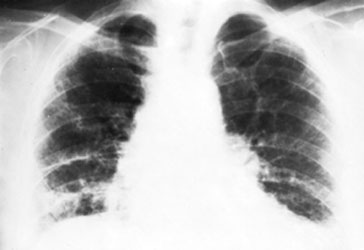
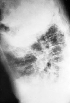
V-17. A 34-year-old man complains of shortness of breath af
ter
minimal exertion. He has no systemic symptoms. He developed a nonproductive
cough 10 months ago. A chest x-ray, which was reportedly normal, was done at
that time. Examination now reveals a respiratory rate of 28 breaths per
minute, and diffuse end-inspiratory crackles are heard over his lower lung
fields. His chest x-rays are shown below. An arterial PO2
measured while the patient is breathing room air is 55 mmHg, and arterial PCO2
is 26 mmHg. Routine blood counts are normal. The next step in his evaluation
should be
The chest x-rays presented show diffuse, severe interstitial infiltrates
without hilar adenopathy. Although sarcoidosis may produce this radiographic
picture, it is also compatible with idiopathic interstitial pneumonitis,
hypersensitivity pneumonitis, collagen vascular disease, inhalation of
inorganic dusts, and many other processes. The degree of respiratory system
dysfunction demonstrated by this patient necessitates rapid evaluation and a
definitive histologic diagnosis so that appropriate therapy can be initiated.
Angiotensin converting enzyme levels, although elevated in many patients with
sarcoidosis, are not sufficiently sensitive or specific to replace tissue
biopsy in the workup of persons with interstitial infiltrates. Although biopsy
of extrapulmonary tissue may demonstrate noncaseating granulomas in patients
with sarcoidosis, such biopsies may be negative in patients with active
disease. A pathologic diagnosis is absolutely required in patients presenting
with interstitial lung disease of uncertain etiology. Fiberoptic bronchoscopy
should be performed to rule out infection or malignancy; an accompanying
transbronchial biopsy may yield a diagnosis about 25 percent of the time.
Bronchoalveolar lavage to assess the degree of inflammation may be helpful in
monitoring disease activity, but its precise role in interstitial lung disease
has not been defined. Despite its relatively low yield, the relatively low
risk makes an attempt at transbronchial biopsy reasonable before definitely
obtaining tissue at open lung biopsy.
V-18. A 23-year-old woman complains of dyspnea and substernal chest
pain on exertion. Evaluation for this complaint 6 months ago included
arterial blood-gas testing, which revealed pH 7.48, PO2
79 mmHg, and PCO2 31 mmHg.
Electrocardiography then showed a right axis deviation. Chest x-ray now
shows enlarged pulmonary arteries but no parenchymal infiltrates, and a lung
perfusion scan reveals subsegmental defects that are thought to have a
"low probability for pulmonary thromboembolism." Echocardiography
demonstrates right heart strain but no evidence of primary cardiac disease.
The most appropriate diagnostic test now would be
(A)
open
lung biopsy
(B)
Holter
monitoring
(C)
right-heart
catheterization
(D)
transbronchial
biopsy
(E)
serum
Alpha1-antitrypsin level
The answer is C.
(Chapter 260.
Rich, Primary Pulmonary Hypertension, in Braunwald E (ed), Heart Disease, 1996.)
The explanation
for the correct response is:
Primary pulmonary hypertension is an uncommon disease that usually affects
young women. Early in the illness affected persons often are diagnosed as
psychoneurotic because of the vague nature of presenting complaints, for
example, dyspnea, chest pain, and evidence of hyperventilation without
hypoxemia on arterial blood-gas testing. However, progression of the disease
leads to syncope in approximately one-half of cases and signs of right heart
failure on physical examination. Chest x-ray typically shows enlarged central
pulmonary arteries with or without attenuation of peripheral markings. The
diagnosis of primary pulmonary hypertension is made by documentation of
elevated pressures by right heart catheterization and exclusion of other
pathologic processes. Lung disease of sufficient severity to cause pulmonary
hypertension would be evident by history and on examination. Major
differential diagnoses include thromboemboli and heart disease; outside the
United States, schistosomiasis and filariasis are common causes of pulmonary
hypertension, and a careful travel history should be taken.
V-19. A 53-year-old man is noted to be tachypneic and confused 48 h
after suffering multiple orthopedic and internal injuries in an automobile
accident. Chest x-ray is interpreted as normal, but arterial blood-gas
values are as follows: pH 7.49, PO2 52
mmHg, and PCO2 30 mmHg. The course of
action most likely to confirm the diagnosis of this man's condition would be
to
The clinical triad of dyspnea, confusion, and petechiae in a person who has
had recent long-bone fractures establishes the diagnosis of fat embolism
syndrome. This disorder, which usually occurs within 48 h of injury, may lead
to respiratory failure and death. Petechiae most often are found across the
neck, in the axillae, and in the conjunctivae; however, their appearance is
often evanescent. No laboratory test is specific for fat embolism.
V-20. All the following statements about obstructive sleep apnea
syndrome are true EXCEPT
(A)
men
are affected more often than women
(B)
systemic
hypertension is a common finding
(C)
alcohol
can be a contributing factor
(D)
estrogens
are frequently useful
(E)
personality
changes may be the presenting complaint
The answer is D.
(Chapter 264.
Fujita, Ear Nose Throat J 72:67, 1993.)
The explanation
for the correct response is:
Obstructive sleep apnea syndrome is a complex entity that involves
intermittent upper-airway obstruction during sleep. Most of the
manifestations, such as hypertension, cor pulmonale, chronic fatigue,
personality changes, and disordered sleep behavior, resolve when obstruction
is bypassed by a tracheostomy or endotracheal tube. Although the syndrome is
more common in men, the prevalence increases in women after menopause. Alcohol
and sedatives can exacerbate ventilatory obstruction by decreasing
upper-airway muscle tone. Treatment of severe obstructive sleep apnea includes
tricyclics to improve upper-airway muscle tone, uvulopalatopharyngoplasty to
create a more spacious airway, continuous nasal positive airway pressure to
prevent muscular collapse, and tracheostomy to bypass the obstruction
completely. Estrogens, which once were thought to be beneficial in improving
respiratory drive, are not now considered a mainstay of treatment.
V-21. A 54-year-old man has a nonproductive cough and exertional
breathlessness. He also notes low-grade fever, malaise, and a weight loss of
7 kg (15 lb) over 6 weeks. His white blood cell count is 13,500/L.
He has a history of mild asthma. A chest x-ray discloses peripheral lung
infiltrates. The most likely diagnosis is
(A)
idiopathic
pulmonary fibrosis
(B)
alveolar
proteinosis
(C)
polymyositis
(D)
chronic
eosinophilic pneumonia
(E)
lymphangiomyomatosis
The answer is D.
(Chapter 253.
Hayakawa, Chest 105:1462, 1992.)
The explanation
for the correct response is:
Chronic eosinophilic pneumonia is an interstitial lung disorder of unknown
cause that produces a systemic illness characterized by fever, weight loss,
and malaise. Although lung biopsy shows an eosinophilic infiltrate involving
both the interstitium and the alveolar space, there may not be an associated
eosinophilia in the peripheral blood. The diagnosis should be suggested by the
"photonegative pulmonary edema" pattern, with central sparing and
nonsegmental, patchy infiltrates in the lung periphery. This disorder often
responds dramatically to corticosteroid therapy. Idiopathic pulmonary fibrosis
and polymyositis produce diffuse reticular, nodular, or reticulonodular
infiltrates on chest x-ray. Alveolar proteinosis is a rare disorder that most
often produces a diffuse air-space filling pattern radiating from hilar
regions on chest x-ray, often with air bronchograms. Alveolar proteinosis does
not cause fever unless it is complicated by an infection such as nocardiosis.
Lymphangiomyomatosis is also rare. It occurs exclusively in women of
childbearing age. The chest x-ray shows reticulonodular infiltration, but the
lungs often appear hyperinflated. Lymphangiomyomatosis is complicated by
pleural effusion and pneumothorax but not by fever.
V-22. Owing to profound hypoxemia, tracheal intubation is performed
on a drowning victim, and mechanical ventilation is begun. Inspired oxygen
concentration is 80%. Initially, the man is agitated and fights the
respirator. Arterial blood gases are obtained and show pH 7.21, PO2
70 mmHg, and PCO2 56 mmHg. The most
appropriate management step at this time would be to
(A)
add
positive end-expiratory pressure (5 cmH2O)
(B)
sedate
the man and control his ventilation
(C)
infuse
sodium bicarbonate intravenously
(D)
raise
the inspired oxygen concentration
(E)
initiate
extracorporeal membrane oxygenation
The answer is B.
(Chapter 266.
Hinson, Annu Rev Med 43:341, 1992.)
The explanation
for the correct response is:
Some persons who become agitated or anxious on a mechanical ventilator receive
inadequate ventilation because they are breathing out of phase with the
machine. The man described in the question has adequate oxygenation; a PO2
of 70 mmHg means that his hemoglobin is more than 90 percent saturated.
However, he is hypoventilating and has developed an acute respiratory
acidosis. Positive end-expiratory pressure (PEEP) improves oxygenation by
raising the lung volume and reducing shunting, but it does not have a large
effect on carbon dioxide clearance. Therefore, the appropriate first step in
management would be to administer a sedative and control the man's ventilation
to reduce arterial PCO2 and raise pH.
V-23. One week after a right total hip replacement a 65-year-old
woman develops the sudden onset of shortness of breath. A workup reveals
normotension, a prominent second heart sound, hypoxemia, sinus tachycardia
with new right axis deviation on the electrocardiogram, and a normal chest
x-ray. Oxygen is administered. Impedance plethysmography is consistent with
a large proximal clot in the left leg. Which of the following would be the
most reasonable next step?
Patients at high risk for thromboembolic disease include those who have had
recent anesthesia, recent childbirth, heart failure, leg fracture, prolonged
bed rest, obesity, estrogen use, or cancer. The clinical scenario presented is
highly consistent with a pulmonary embolism arising from venous thrombosis of
a proximal lower extremity in a postoperative patient. While the
electrocardiogram is usually normal except for sinus tachycardia, the finding
of new right-sided heart strain is compatible with a significant pulmonary
embolus. The positive impedance plethysmogram for an above-the-knee venous
thrombosis obviates the need for additional diagnostic testing. The patient
must receive antithrombotic therapy (heparin) in an attempt to inhibit clot
growth, promote resolution, and prevent recurrence. Warfarin requires several
days to achieve anticoagulation and is therefore not appropriate in the acute
setting. While thrombolytic therapy can clearly hasten the resolution of
thrombi and may be appropriate for large, deep venous thromboses and pulmonary
embolisms large enough to cause hypotension, its role in altering the natural
history of this disorder has not been defined. Furthermore, recent surgery is
a contraindication to the use of thrombolytic agents, which, even in the case
of more specific newer agents such as tissue plasminogen activator, carry
significant hemorrhagic risk. Lower-molecular-weight heparins (e.g.,
enoxaporin) have several potential advantages, including a longer half-life
and a more predictable dose response, compared with unfractioned heparin;
however, this newer agent has not received FDA approval for the treatment of
pulmonary embolism or deep venous thrombosis.
V-24. Which of the following is LEAST likely to be associated with
cystic fibrosis?
Although the majority of patients with cystic fibrosis are diagnosed in
childhood, a significant number of patients are not identified until their
late teens, twenties, or even thirties. Accurate diagnosis requires that the
sweat chloride test be given to all patients with clinical features of cystic
fibrosis. Airway obstruction resulting from bronchiectasis is associated with
sinusitis and infertility in males with both cystic fibrosis and the immotile
cilia syndrome, but only males with immotile cilia have Kartagener's syndrome
(bronchiectasis, sinusitis, and dextrocardia). Patients with cystic fibrosis
may have any of several gastrointestinal manifestations including obstruction,
intussusception, fecal impaction, volvulus, portal hypertension, and
steatorrhea. Steatorrhea is a manifestation of pancreatic insufficiency.
Nearly all patients with cystic fibrosis display clubbing.
V-25. Expected results of the pulmonary function testing of the man
described above would include
The man described in the question presents with physical signs (pursed lip
breathing, chest hyperexpansion) and radiographic evidence (flattened
diaphragms, attenuated markings) suggestive of obstructive lung disease with
loss of lung tissue. Reduced expiratory air-flow rates are produced by
narrowing of airways (e.g., in asthma), loss of airways (e.g., in
bronchiolitis obliterans), or loss of elastic tissue (e.g., in emphysema).
Pathophysiologically, these conditions cause increased resistance as airways
are narrowed or collapse as well as decreased driving pressure that represents
loss of elastic recoil. Air trapping and reduced lung recoil lead to an
increase in both total lung capacity (TLC) and functional residual capacity (FRC),
which is the volume at which the tendency of the lung to recoil inward is just
balanced by the tendency of the chest to recoil outward. Although TLC is
increased, vital capacity, the maximum amount of gas that can be exhaled from
the lungs with a single breath, is reduced owing to the great increase in
residual volume produced by gas trapping. Not only is vital capacity reduced,
it takes longer to empty the lungs; thus, forced expiratory volume in 1 s (FEV1)
is reduced as a percentage of vital capacity. When alveolar capillaries are
destroyed by emphysema, the diffusing capacity, which reflects in part the
surface area of alveolar membrane available for gas exchange, is reduced.
V-26. Initial laboratory assessment of the man described above
should include all the following EXCEPT
To establish baseline information in persons who have emphysema, spirometry
should be performed, and for persons with significant complaints or physical
findings, arterial blood gases also should be checked. Although cigarette
smoking accounts for the vast majority of cases of emphysema, a small
percentage of persons who develop this illness have had no exposure to tobacco
products. A subset of this nonsmoking, emphysematous population is deficient
in 1-antitrypsin,
which is a protease inhibitor that normally is found in the serum. It is
currently believed that release of proteolytic enzymes from inflammatory cells
accounts for the lung destruction that typifies emphysema, and 1-antitrypsin
deficiency, a familial disorder, the genotype of which is acid starch gel and
immunoelectrophoresis, permits this destruction to occur unimpeded. Exercise
testing is not necessary as an initial screening test for emphysema but should
be considered before oxygen therapy is prescribed. A male who has
emphysematous respiratory failure, gives no history of respiratory infections,
and has children would not have cystic fibrosis (affected men are sterile);
therefore, a sweat chloride test would not be a useful procedure.
V-27. Correct statements concerning the pathogenesis of 1-antitrypsin
deficiency include all the following EXCEPT
(A)
emphysema
results from an inability to inhibit alveolar destruction by
neutrophils
(B)
clinical
deficiency of 1-antitrypsin
usually results from one of several missense mutations that cause a
truncated mRNA
(C)
the
disease is inherited in a dominant fashion
(D)
mutations
of the 1-antitrypsin
gene of the S type produce less severe emphysema than do mutations
of the Z type
(E)
treatment
with purified 1-antitrypsin
can raise the serum level to that associated with lung protection
Reduced serum levels of the antiprotease &agrl1-antitrypsin,
which is synthesized primarily in the liver, are associated with an inability
to control the alveolar-damaging effects of neutrophil elastase and clinical
emphysema. 1-Antitrypsin
is encoded by a 7-exon gene spanning 12.2 kilobases on chromosome 14. Common
disease-producing mutations of the normal M gene are the Z type, in which a
single amino-acid substitution results in a hyperaggregative, improperly
processed protein, and the S type, which results in a product with a shortened
half-life and also is due to a single amino-acid change. Heterozygotes (either
M2 or M5) appear to have sufficient, albeit reduced, levels of 1-antitrypsin
to prevent severe lung damage. Since the S type produces less clinical
antiprotease "deficiency," the pulmonary disease produced in SS
homozygotes is much less severe than that seen in patients whose genotype is
ZZ. Intravenous administration of normal human purified 1-antitrypsin
can increase serum levels to a point at which sufficient antiprotease activity
is provided to protect alveoli from elastase-induced damage.
V-28. To decrease the likelihood of drug toxicity, the theophylline
dose should be reduced in a patient with asthma in each of the following
circumstances EXCEPT
Although inhaled sympathomimetics are now considered the first-choice
treatment for acute asthmatic attacks, methylxanthines such as theophylline
are effective bronchodilators and continue to be used extensively in this
disorder. The therapeutic plasma concentration is 10 to 20 mg/mL, but the dose
required to achieve these levels varies widely, depending on the clinical
situation. The theophylline dose should be reduced in any condition in which
the clearance of this drug is significantly impaired, such as in the very
young, the elderly, and those with liver or cardiac dysfunction. Many drugs
interfere with the metabolism of theophylline. Some commonly used agents,
including allopurinol, propranolol, cimetidine, and erythromycin, interfere
with theophylline clearance and thus lead to increased levels of this
methylxanthine. Drugs that activate hepatic microsomal enzymes, such as
cigarettes, marijuana, phenobarbital, and phenytoin, may lower theophylline
levels.
V-29. Known consequences of asbestos exposure include all of the
following EXCEPT
Inhalation of asbestos fibers for 10 years or more may lead to interstitial
fibrosis that typically begins in the lower lobes and later spreads to the
middle and upper lung fields. This fibrosis is associated with a restrictive
pattern on pulmonary function testing. The chest x-ray shows linear densities,
thickening or calcification of the pleura (pleural plaques), and, in severe
cases, honeycombing. Exposure to asbestos also may cause exudative pleural
effusions. These effusions are often bloodstained and may be painful. The
diagnosis may be elusive if a careful occupational exposure is not obtained.
These effusions are benign, but affected persons may later sustain malignant
mesotheliomas of the pleura or peritoneum. Unlike pulmonary fibrosis, pleural
effusions and mesotheliomas may develop after brief exposures to asbestos,
often exposures of 1 to 2 years. Mesotheliomas are not associated with
cigarette smoking, but the combination of exposure to asbestos and cigarette
smoking has a multiplicative effect on the risk of development of lung cancer.
Exposure to asbestos increases the risk for both adenocarcinoma and squamous
cell (but not small cell) carcinoma of the lung; this suggests that lung
cancer screening may be useful in selected individuals.
V-30. In which of the following clinical circumstances would it be
appropriate to use a rigid bronchoscope instead of a flexible fiberoptic
bronchoscope?
(A)
A
22-year-old male with known HIV infection who complains of shortness
of breath and has diffuse interstitial infiltrates on chest x-ray
(B)
A
65-year-old male with a long history of smoking who has shortness of
breath and right upper lobe collapse
(C)
A
33-year-old female with a history of acute myeloid leukemia
complaining of severe dyspnea who is currently 4 months after an
allogeneic bone marrow transplant and has a reticulonodular
pulmonary infiltrate
(D)
A
50-year-old female with a heavy smoking history who currently
complains of intermittent hemoptysis
(E)
A
28-year-old male with a history of acute myeloid leukemia who is
currently 30 days after an allogeneic bone marrow transplant with a
significant pulmonary hemorrhage and bilateral alveolar infiltrates
on chest x-ray
The flexible fiberoptic bronchoscope is ideal for many clinical situations,
including those designed to determine many endobronchial pathologic states,
including tumors, granulomas, bronchitis, foreign bodies, and sites of
bleeding. Washing or infiltration of a higher volume of sterile saline (bronchoalveolar
lavage) can be used to detect abnormal cells or recover pathogens such as P.
carinii in patients with HIV infection. Brushing or biopsy at the surface
of an endobronchial lesion can enhance the recovery of cellular material or
tissue that can be very helpful in detecting neoplasms or infection. Pulmonary
hemorrhage can complicate a transbronchial biopsy if the patient is at risk
for a bleeding diathesis. Pneumothorax can occur if the forceps are too close
to the pleural surface. Because a rigid bronchoscope has a larger suction
channel and allows ventilation, this instrument is still useful for the
retrieval of foreign bodies and the suctioning of massive hemorrhages.
V-31. The most common initial symptom of byssinosis is
Pulmonary disease secondary to cotton dust exposure is one of the most common
occupational lung diseases. During the production of yarn for cotton, linen,
and rope making, exposure to cotton, flax, or hemp produces a host of
respiratory symptoms. Exposure to cotton dust (byssinosis) is characterized as
chest tightness toward the end of the first day of the workweek. Such symptoms
are associated with a significant drop in the forced expiratory volume during
the workday. Although most workers have no recurrence after the workweek, up
to 25 percent may have a progressive symptom complex consisting of recurrent
chest tightness, eventually leading to an obstructive pattern on pulmonary
function testing. The chest tightness appears to be due to bronchospasm, which
may be reversible with bronchodilators. The best treatment is a reduction of
dust exposure.
V-32. A 27-year-old female with a history of common variable
immunodeficiency has had many upper and lower respiratory tract infections.
She now presents with a third episode of recurrent cough and copious
purulent sputum production, which is sometimes blood-tinged. She is afebrile,
and her pulmonary exam is normal. Chest radiography reveals the presence of
several parallel linear opacities and a few ringlike shadows. The diagnosis
that most likely accounts for this patient's symptoms is
Bronchiectasis represents an abnormal permanent dilation of the bronchi,
typically on the basis of chronic destruction and inflammation caused by
repetitive infection or other chronic insults. Many infectious agents,
including adenovirus, influenza virus, Staphyloccoccus aureus,
tuberculosis, and anaerobic infection, can each predispose a patient to the
bronchiectatic state. Problems of primary immune defenses such as
immunoglobulin deficiency, primary cilliary disorders, and cystic fibrosis
also can produce dilated bronchi. This anatomic problem leads to recurring
cough and purulent sputum production, frequently associated with hemoptysis
resulting from friable, inflamed airway mucosa. Repetitive bronchiectatic
episodes tend to produce increased problems. Physical examination is
nonspecific; however, the chest radiograph is frequently abnormal, and the
findings may include cystic spaces caused by saccular bronchiectasis or the
so-called tram track (parallel linear shadows) or rings (produced if the
inflamed thickened airways are seen in cross section). Treatment requires
elimination of the underlying problem (e.g., by immunoglobulin infusions),
improved clearance of tracheobronchial secretions, control of infection, and
the use of bronchodilators to reverse airflow obstruction.
V-33. In which of the following situations would single-lung
transplantation be contraindicated?
(A)
A
48-year-old male with chronic obstructive pulmonary disease and an
FEV1 of 20 percent of the predicted value
(B)
A
50-year-old male with idiopthic pulmonary fibrosis,resting hypoxia,
and a total lung capacity of 50 percent of the predicted value.
(C)
A
23-year-old female with primary pulmonary hypertension with a mean
pulmonary artery pressure of 70 mmHg
(D)
A
23-year-old female with cystic fibrosis and an FEV1 of 20
percent of the predicted value
(E)
A
25-year-old male with an 1-antitrypsin
deficiency and resting hypoxia
Emphysema, either smoking-induced or resulting from 1-antitrypsin
deficiency, is the indication for almost 50 percent of all single-lung
transplants. It is important to determine the optimal lung transplant window
in which the patient has severe limitations secondary to his or her disease
but has not passed the point at which lung transplant surgery would be
dangerously complicated. Life expectancy of less than 2 years and severe
obstructive lung disease (FEV1 of less than 30 percent of the
predicted value) are required. Patients with pulmonary fibrosis must have
profound impairment of total lung capacity and resting hypoxia; those with
pulmonary hypertension need to be severely functionally limited and have
pulmonary artery pressures greater than 50 mmHg. Patients with cystic fibrosis
must also have severe limitations in expiratory capacity as well as
abnormalities in arterial blood gases. They are not candidates for single-lung
transplantation because of the risk of disseminated infection. Instead, they
require a bilateral lung transplant or a related donor bilobe transplant to
reduce the likelihood of disseminated infection. In each of the other
situations, single-lung transplantation can be performed with acceptable risk
and tangible benefit for the patient.
V-34. A 50-year-old male chronic alcoholic presents with a 2-week
history of fever, night sweats, cough, productive sputum, and pleuritic
chest pain. The patient has had a recent negative HIV test and has no other
medical problems. Chest x-ray reveals a 3-cm cavitary lesion in the
posterior segment of the left lower lobe. This cavity contains an air-fluid
level. Which of the following is the most likely etiologic agent?
Microbial pathogens may enter the lung by several routes, including aspiration
of organisms that colonize the oropharynx, inhalation of infectious aerosols,
direct inoculation (e.g., from tracheal intubation or stab wounds), and
hematogenous dissemination from an extrapulmonary site. In a chronic
alcoholic, presumably with a higher likelihood of aspiration of oral contents,
one must seriously consider the presentation of an aspiration pneumonia caused
by anaerobic oral flora. Moreover, certain oral anaerobes, such as S.
aureus, S. pneumoniae serotype III, aerobic gram-negative bacilli,
oral anaerobes, M. tuberculosis, and fungi, produce tissue necrosis and
pulmonary cavities. H. influenzae, M. pneumoniae, and other type
serotypes of S. pneumoniae are less likely to cause cavities. Cavities
associated with M. tuberculosis do have air-fluid levels but are
typically in the upper lobe as a result of the fact that they require high
oxygen tension for optimal growth. By contrast, a cavity containing an
air-fluid level in a dependent, fully ventilated, fully draining
bronchopulmonary segment suggests that the culprit is an oral anaerobe. One
particular oral anaerobe, Actinomyces spp., can produce a chronic
fibrotic necrotizing process that can cross tissue planes to involve the
pleural space, ribs, vertebrae, and subcutaneous tissue with the eventual
discharge of sulfur granules.
V-35. A 55-year-old male presents with several months of dyspnea
and a nonproductive cough. Physical examination reveals dry crackles at both
lung bases. Chest radiography and high-resolution computed tomography reveal
a bibasilar reticular nodular pattern in the lung fields. Spirometry reveals
reductions in total lung capacity, vital capacity, and residual volume. The
carbon monoxide diffusion capacity is reduced to 35 percent of normal.
Resting arterial hypoxemia is demonstrated on arterial blood-gas testing.
Transbronchial biopsy results reveal an increase in inflammatory cells on
the alveolar surface, predominantly macrophages, as well as diffuse
intraalveolar fibrosis. The mainstay of therapy at this point would be
(A)
oral
prednisone
(B)
oral
cyclophosphamide
(C)
4-week
course of oral azithromycin
(D)
lung
transplantation
(E)
bronchodialator
therapy
The answer is A.
(Chapter 259.
Hunninghack, Am J Respir Crit Care Med 151:915, 1995.)
The explanation
for the correct response is:
This patient presents with the classic history, physical findings, and
pathologic findings consistent with idiopathic pulmonary fibrosis. This is an
unrelenting interstitial lung disease which produces scarring and ablation of
alveoli with a concomitant restrictive lung pattern. If progressive and/or
unresponsive to therapy, the disease will lead to progressive loss of
pulmonary function and ultimately to right-sided heart failure. The primary
cause is unknown, although the pathophysiology clearly involves the activation
of alveolar macrophages, with secondary cytokine release causing alveolar
damage with associated fibrosis. Once the diagnosis is made, even at an
advanced stage, a trial of glucocorticoids is indicated. High-dose steroid
therapy for 8 weeks usually is initiated with gradual tapering if objective
improvement is noted. Cyclophosphamide may be useful in patients who are
unresponsive to glucocorticoids. The dose of cyclophosphamide should be
titrated to ensure that the total neutrophil count does not drop below 1000/L.
Drugs such as penicillamine, cyclosporine, and colchicine, each of which could
potentially play a role in inhibiting macrophage-produced growth factors,
remain investigative. Although measures to prevent progressive restrictive
physiology include diuretics and oxygen for congestive heart failure, rapid
treatment of infection, and the routine use of prophylactic pneumococcal
influenza vaccine, in the situation of a patient who is in otherwise good
medical condition but has profoundly deranged pulmonary physiology secondary
to fibrosis, lung transplantation should be considered.
V-36. A 19-year-old normal nonsmoking female has a moderately
severe pulmonary embolism while on oral contraceptive pills. Which of the
following is the most likely predisposing factor?
(A)
Abnormal
factor V
(B)
Abnormal
protein C
(C)
Diminished
protein C level
(D)
Diminished
protein S level
(E)
Diminished
antithromin III level
The answer is A.
(Chapter 261.
Ridker, N Engl J Med 332:912, 1995.)
The explanation
for the correct response is:
Many patients who develop pulmonary thromboembolism have an underlying
inherited predisposition that remains clinically silent until they are
subjected to an additional stress, such as the use of oral contraceptive
pills, surgery, or pregnancy. The most frequently inherited predisposition to
thrombosis is so-called activated protein C resistance. The inability of a
normal protein C to carry out its anticoagulant function is due to a missense
mutation in the gene coding for factor V in the coagulation cascade. This
mutation, which results in the substitution of a glutamine for an arginine
residue in position 506 of the factor V molecule, is designated the factor V
Leiden gene. Based on the Physicians Health Study, about 3 percent of healthy
male physicians carry this particular missense mutation. Carriers are clearly
at an increased risk for deep venous thrombosis and also for recurrence after
the discontinuation of coumadin. The allelic frequency of factor V Leiden is
more common than are all other identified inherited hypercoagulable states
combined, including deficiencies of protein C, protein S, and antithrombin III
and disorders of plasminogen.
V-37. A 65-year-old male presents for an evaluation because he is
"feeling poorly." Symptoms include morning headache and poor sleep
quality. He is quite tired during the day and frequently falls asleep while
he reads or watches television. Physical examination reveals a ruddy
complexion but is otherwise unremarkable. Laboratory examination is normal
except for elevations in hematocrit and plasma HCO3-
concentration. Polysomnography demonstrates a decreased ventilatory response
to hypercapnia and many episodes of central apnea (no diaphragmatic activity
is noted). The maximum respiratory pressure that he generates against an
occluded airway is normal. Spirometry and blood gases are normal. Of the
following, which is the most likely cause of the patient's problem?
The hypoventilation syndromes are defined as disorders which yield to
hypercapnia (usually a PaCO2 in the range of
50 to 80 mmHg). The clinical manifestations of such a syndrome include
respiratory acidosis with a compensatory rise in the plasma bicarbonate
concentration and a decrease in chloride concentration. The hypercapnia leads
to an obligatory decrease in the arterial oxygen tension with subsequent
cyanosis and secondary polycythemia. Moreover, chronic hypercapnia can induce
pulmonary vasoconstriction which can eventually lead to right ventricular
failure. Problems of hypoventilation normally are exacerbated at night,
leading to worsened hypercapnia during sleep with subsequent morning headache
and daytime somnolence with eventual intellectual impairment. A polysomnogram
can distinguish whether the defect is in the metabolic control system (chemoreceptors
or brainstem initiating neurons) versus the neuromuscular system (brainstem
neurons, spinal cord, respiratory nerves) or in the ventilatory apparatus
(e.g., chest wall, lungs, airways) itself. Those with a central problem such
as higher CNS malfunction or chemoreceptor insufficiency have normal
spirometry, blood gases, and tests of voluntary hyperventilation. However,
they would have a markedly blunted response to hypoxia or hypercapnia.
Respiratory neuromuscular dysfunction that produces a decrease in the ability
to exhale would lead to an abnormal spirometric evaluation and also would make
it impossible for the patient to generate normal inspiratory and expiratory
muscle pressures against a closed airway. Those with problems with the
ventilatory apparatus, such as those which typically occur in obstructive
sleep apnea, would be able to generate adequate expiratory and inspiratory
muscle pressures and generally would have markedly abnormal blood gases (e.g.,
a widened arterial alveolar oxygen tension gradient).
V-38. All the following are typical manifestations of chronic
hyperventilation EXCEPT
In all cases of chronic hyperventilation the mechanism involves an increase in
respiratory drive that may well be physiologic (e.g., chronic hypoxemia) but
can be detrimental because of the ensuing alkalemia. This disturbance in blood
pH can produce neurologic symptoms such as dizziness, syncope, and seizure
activity caused by cerebral vasoconstriction. The neuromuscular side effects
of chronic alkalemia can include paresthesia, muscle weakness (from
hypophosphatemia), and hypocalcemia-induced carpopedal spasm tetany. The
primary respiratory alkalosis can lead to central sleep apnea. The disorders
that most frequently lead to unexplained hyperventilation are pulmonary
vascular diseases such as chronic thromboembolism and anxiety. In patients who
have symptoms clearly secondary to hyperventilation, inhalation of a low
concentration of carbon dioxide can be helpful.
V-39. Which of the following circumstances leading to the acute
respiratory distress syndrome and the necessity for mechanical ventilation
would have the best prognosis?
(A)
A
33-year-old male with a heroin overdose
(B)
A
68-year-old male with an acute myocardial infarction and 2 h of
hypotension
(C)
A
25-year-old male poststatus a gunshot wound, major volume loss,
hypotension, and acute renal failure
(D)
A
21-year-old female with acute myeloid leukemia with gram-negative
sepsis during induction therapy
(E)
A
45-year-old fireman with severe smoke inhalation injury and arterial
PaO2 of 60 mmHg despite 100 percent FIO2
The answer is A.
(Chapter 265.
Milberg, JAMA 273:306, 1995.)
The explanation
for the correct response is:
The acute respiratory distress syndrome is a condition characterized by
hypoxemic respiratory failure resulting from noncardiogenic pulmonary edema.
Whatever the initial insult, the final common pathway of diffuse lung injury
represents a cascade of cellular events that are associated with cytokine
production by inflammatory cell activation and the production of inflammatory
mediators which damage alveolar and pulmonary endothelial cells, leading to
increased vascular permeability and loss of surfactant production by type II
pneumocytes. When the injury is severe, mechanical ventilation is required.
Despite the critical involvement of the inflammatory response in the
generation of acute respiratory distress syndrome, glucocorticoids have shown
no benefit in the treatment of this condition, except in childhood
meningococcemia and P. carinii pneumonia. Although mortality rates for
acute respiratory distress syndrome range from 50 to 70 percent, it is likely
that improvements in therapy are reducing this dire outcome. Age greater than
65 years, multiorgan system failure, sepsis, and severe gas exchange
disturbances are all negative prognostic factors. However, those with
uncomplicated overdoses tend to have a relatively improved outcome. Those who
survive are likely to return to the preexisting level of function.
V-40. All the following represent strategies to deal with the
complications of mechanical ventilation EXCEPT
(A)
thoracostomy
placement for barotrauma-induced pneumothorax
Enterotracheal intubation with positive-pressure mechanical ventilation may
save lives in multiple settings associated with respiratory failure. This
highly invasive technique has both direct and indirect effects on many organ
systems. Lung complications include barotrauma (especially in the setting of
the use of high levels of positive end-expiratory pressure), nosocomial
pneumonia, oxygen toxicity, tracheal stenosis, and deconditioning of
respiratory muscles. If a clinically significant pneumothorax occurs (e.g.,
associated with hypoxemia, decreased lung compliance, and hypotension),
placement of a thoracostomy tube is required. While nosocomial pneumonia is
common in patients intubated for more than 72 h because of aspiration of oral
pharyngeal contents caused by leaks around the endotracheal tube cuff, the use
of prophylactic antibiotics is not indicated. Moreover, it can be difficult to
distinguish between colonization and true infection, since virtually all
ventilated patients can be shown to have potentially pathogenic bacteria in
the lower respiratory tract. Oxygen toxicity, which may result from the effect
of oxygen free radicals on lung tissues, needs to be managed with the
conservative use of inspired oxygen tensions. Right-sided heart hemodynamic
monitoring often is required to provide optimal levels of intravascular volume
replacement in these patients. It is important to maintain adequate venous
return, but increased lung water also must be avoided. Mild to moderate
cholestasis and stress ulceration are two important gastrointestinal effects
of mechanical ventilation. If the total bilirubin values are greater than 4.0
mg/dL, it is likely that a cause of liver damage other than intubation is
operative. Prophylactic therapy with sucralfate or an H2-receptor
antagonist is helpful in preventing stress-related ulcers in intubated
patients.
V-41. A 65-year-old man with chronic bronchitis presented to the
emergency room 2 weeks ago with acute respiratory failure. He was intubated
and treated with diuretics and antibiotics. However, after apparent
improvement during a 1-week stay in the intensive care unit on mechanical
ventilation, he has failed three attempts at being weaned from the
ventilator. All the following factors could account for the difficulty in
removing this patient from the ventilator EXCEPT
There are many reasons why patients fail removal from assisted ventilation.
Sedatives, which are commonly prescribed earlier in the hospital stay for
agitation or sleep, may not have been discontinued or metabolized and could
contribute to impairment of respiratory drive. Persistent secretions that
could be removed by suctioning also might contribute to the problem. A very
important issue is maintenance of a continued drive to breathe. The central
respiratory centers are sensitive to blood pH and will promote ventilation
when sufficient acidosis (greater than that normally noted in a bronchitic
patient with chronic CO2 retention) ensues. Thus, creation of
metabolic alkalosis with diuretic therapy or running the assisted minute
ventilation too high (i.e., lower PCO2 and higher pH than normal
for the patient) could account for failed extubation. Neuromuscular weakness
caused by diuretic-induced hypokalemia, malnutrition, and occult
hypothyroidism are potential factors leading to difficulty in independent
ventilation and trouble in weaning.
V-42. Hypoxemia occurring after pulmonary thromboembolism can
result from
(A)
lowered
mixed venous PO2 resulting from heart failure
(B)
perfusion
of atelectatic areas
(C)
increased
dead-space ventilation in the area of vascular occlusion
(D)
perfusion
of areas poorly ventilated because of airway constriction
(E)
inadequate
time for oxygen diffusion secondary to a reduction in the capillary
bed
Hypoxemia occurs commonly after massive pulmonary thromboembolism, although
normal arterial oxygen tension does not exclude the diagnosis. The most
important mechanism producing hypoxemia in this setting is an increase in
venous admixture caused by continued perfusion of poorly ventilated areas.
Ventilation may be decreased by atelectasis or by airway constriction in
response to the release of bronchoactive mediators. A fall in cardiac output
that produces a low mixed venous PO2 can
increase the effect of venous admixture. Increased dead-space ventilation
would not be a cause of hypoxemia.
V-43. Which of the following conditions would be likely to result
in an increased residual volume on plethysmographic pulmonary function
testing?
The volume remaining in the lungs at the conclusion of a complete forced
expiration is termed the residual volume and can be determined either
by the body plethysmography or by helium dilution methods. At the residual
volume there is a balance between the intrinsic outward recoil of the chest
wall and the force maintained by the respiratory muscles to decrease lung
volumes further. Therefore, increases in the residual volume can result from
the functionally weak musculature of the chest wall that might be observed in
neuromuscular disorders that affect the ability to expire forcefully (Guillain-Barré
syndrome, muscular dystrophies, cervical spine injury). Furthermore, diseased
airways will collapse at low lung volumes, preventing further emptying and
also producing an abnormally high residual volume. Thus, any condition in
which airway obstruction plays a major role (chronic bronchitis, emphysema,
asthma, cystic fibrosis) may be associated with increased residual volume. By
contrast, pulmonary parenchymal disease (e.g., sarcoidosis) produces normal
expiration and reduced lung volumes. If inspiratory dysfunction is the primary
chest wall problem (as in kyphoscoliosis and obesity), residual volume will be
relatively unaffected or slightly decreased.
V-44. A 51-year-old man develops pancreatitis associated with the
passage of a gallstone. His treatment includes meperidine and intravenous
normal saline. Two days later he becomes anxious, tachypneic, and short of
breath. An emergency chest x-ray demonstrates diffuse, bilateral
interstitial and alveolar infiltrates. A year ago he suffered a myocardial
infarction, but since then he has had no evidence of congestive heart
failure. In this case, adult respiratory distress syndrome can be
distinguished from cardiogenic pulmonary edema by
(A)
measurement
of lung water
(B)
measurement
of protein concentraton in edema fluid
(C)
measurement
of pulmonary artery wedge pressure
(D)
measurement
of lung compliance
(E)
calculation
of the alveolar-arterial PO2 difference
The answer is B,C.
(Chapter 265.
Kolleff, N Engl J Med 332:27, 1995.)
The explanation
for the correct response is:
The adult respiratory distress syndrome (ARDS) is a clinical triad of
hypoxemia, diffuse lung infiltrates, and reduced lung compliance not
attributable to congestive cardiac failure. This syndrome's many causes
suggest its complex pathogenesis. However, the pathologic outcome is the same:
an increase in lung water caused by an increase in alveolar capillary
permeability. This noncardiogenic pulmonary edema is identical to congestive
cardiac pulmonary edema in its effect on the mechanical properties of the lung
and on gas exchange. Just as in cardiac pulmonary edema, the increase in lung
water associated with ARDS produces interstitial edema and alveolar collapse,
and so the affected lung becomes stiff and the alveolar-arterial oxygen
tension difference widens. Unlike cardiac edema, however, the increase in lung
water in ARDS occurs as a result of an increase in alveolar capillary
permeability and is not due to an increase in hydrostatic forces. Edema fluid
in ARDS therefore often contains macromolecules (such as serum proteins), and
measurement of pulmonary artery wedge pressure is normal or low. In clinical
practice, determination of pulmonary artery wedge pressure is the most helpful
discriminant between ARDS and cardiac failure.
 kat/L
(90 U/L). Cell count reveals 1000 red cells per microliter and 1000 white
cells per microliter (differential: 50 percent neutrophils, 25 percent
lymphocytes, and 25 percent monocytes). A ventilation-perfusion lung scan is
indeterminate on the right side because of the large effusion, but there are
no ventilation-perfusion mismatches elsewhere. The next most appropriate
step would be
kat/L
(90 U/L). Cell count reveals 1000 red cells per microliter and 1000 white
cells per microliter (differential: 50 percent neutrophils, 25 percent
lymphocytes, and 25 percent monocytes). A ventilation-perfusion lung scan is
indeterminate on the right side because of the large effusion, but there are
no ventilation-perfusion mismatches elsewhere. The next most appropriate
step would be
 ter
minimal exertion. He has no systemic symptoms. He developed a nonproductive
cough 10 months ago. A chest x-ray, which was reportedly normal, was done at
that time. Examination now reveals a respiratory rate of 28 breaths per
minute, and diffuse end-inspiratory crackles are heard over his lower lung
fields. His chest x-rays are shown below. An arterial PO2
measured while the patient is breathing room air is 55 mmHg, and arterial PCO2
is 26 mmHg. Routine blood counts are normal. The next step in his evaluation
should be
ter
minimal exertion. He has no systemic symptoms. He developed a nonproductive
cough 10 months ago. A chest x-ray, which was reportedly normal, was done at
that time. Examination now reveals a respiratory rate of 28 breaths per
minute, and diffuse end-inspiratory crackles are heard over his lower lung
fields. His chest x-rays are shown below. An arterial PO2
measured while the patient is breathing room air is 55 mmHg, and arterial PCO2
is 26 mmHg. Routine blood counts are normal. The next step in his evaluation
should be